Detecting Gray Matter Maturation via Tensor-based Surface Morphometry
Moo K. Chung

 ,
Keith J. Worsley
,
Keith J. Worsley 1,
S. Robbins1, Tomás Paus1, Jay N. Giedd2,
Judith L. Rapoport2, Alan C. Evans1
1,
S. Robbins1, Tomás Paus1, Jay N. Giedd2,
Judith L. Rapoport2, Alan C. Evans1
Department of Statistics
Department of
Biostatistis and Medical Informatics
W.M. Keck laboratory
for functional brain imaging and behavior, University of Wisconsin-Madison
Department of Mathematics
and Statistics, McGill University
1Montreal Neurological Institute
2Child Psychiatry Branch, National Institute of Mental
Health
Abstract
We present a unified computational approach to tensor-based surface
morphometry in detecting the gray matter growth patterns for 28 children
and young adults aged between 12 and 16. The gray matter has the topology
of a 2D highly convoluted thin sheet. As the brain develops over time,
the cortical surface area, thickness, curvatures and the total gray matter
volume change. It is highly likely that such age-related surface deformations
are not uniform. By measuring how such surface metrics change over time,
the regions of the most rapid structural changes can be detected.
Methods
We used anatomic segmentation using proximities (ASP) method (McDonalds
et al, 2001) to generate both outer and inner surface meshes from classified
MRIs. Then the surface meshes were parameterized by local quadratic polynomials
(Chung et al, 2003). The cortical surface deformation was modeled as the
boundary of ulticomponent fluids (Drew, 1991). Using the same stochastic
assumption on the deformation field used in Chung et al. (2001), the distributions
of area dilatation rate, cortical thickness and curvature changes are derived
(Chung et al, 2003). To increase the signal to noise ratio, diffusion smoothing
(Andrade,2001; Chung et al. 2003) has been developed and applied to surface
data. Afterwards, statistical inference on the cortical surface is performed
via random fields theory (Worsley et al., 1994).
Results
It is found that the total cortical surface area and gray matter volume
shrinks, while the cortical thickness and curvature tends to increase between
ages 12 and 16 with a highly localized area of cortical thickening and
surface area shrinking found in the superior frontal sulcus at the same
time. It seems that the increase in thickness and decrease in the superior
frontal sulcus area are causing increased folding in the middle and superior
frontal gyri (see Figure 1.). Because our technique is based on coordinate-invariant
tensor geometry, artificial surface flattening (Angenent et al., 1999),
which can destroy the inherent geometrical structures of the cortical surface,
has been avoided.
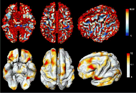 |
| Figure 1 Top: Bending energy computed on the inner cortical
surface of a 14 year old subject. Bottom: t map showing the regions of
curvature increase between ages 12 and 16. Most of curvature increase occurs
on gyri. |
References
Andrade, A., Kherif, F., Mangin, J., Worsley, K.J., Paradis, A., Simons,
O., Dehaene, S., Le Bihan, D., Poline J-B. (2001) Detection of fMRI activation
using cortical surface mapping, Human Brain Mapping, 12:79-93.
Angenent, S., Hacker, S., Tannenbaum, A., Kikinis, R. (1999). On the
Laplace-Beltrami Operator and Brain Surface Flattening, IEEE Transactions
on Medical Imaging 18:700-711.
Chung, M.K., Worsley, K.J., Paus, T., Cherif, D.L., Collins, C., Giedd
J., Rapoport, J.L., Evans, A.C. (2001) A unified statistical approach to
deformation-based morphometry, NeuroImage, 14:595-606.
Chung, M.K., Worsley, Robbins, S, Paus, T., Taylor, J., Giedd, J.N.,
Rapoport, J.L., Evans, A.C. (2003) Deformation-based surface morphometry,
with an application to gray matter deformation, NeuroImage, in press. http://www.stat.wisc.edu/~mchung/papers/surface_ni.pdf
MacDonalds, J.D., Kabani, N., Avis, D., Evans, A.C. (2001) Automated
3-D extraction of inner and outer surfaces of cerebral cortex from MRI,
NeuroImage, 12:340-356.
Worsley, K.J., Marrett, S., Neelin, P., Vandal, A.C., Friston, K.J.,
Evans, A.C. (1996) A unified statistical approach for determining significant
signals in images of cerebral activation, Human Brain Mapping, 4:58-73.